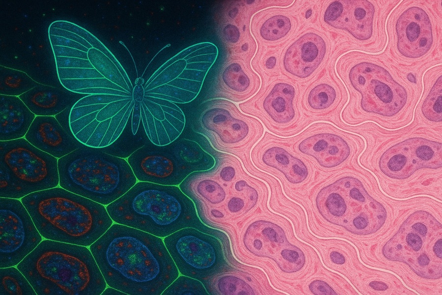
The Immunity Butterfly Effect
How spatial biology and causal AI are redefining drug discovery
Founder and operator. Built Voyant Bio, a spatial biology and AI platform that maps immune cell interactions in cancer to design biologics.
I've operated as scientist, founder, and investor at once: building an AI drug discovery platform, generating a therapeutic candidate, and evaluating other founders' platforms from the other side of the table.
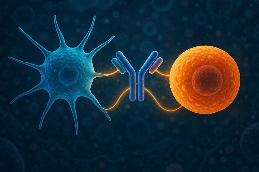
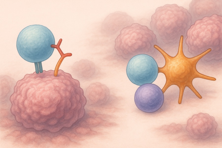
My latest research, published in Nature Medicine, found that response to PD-1 blockade depends on a specific spatial arrangement: dendritic cells organizing CXCL13⁺ CD4⁺ T helper cells and progenitor CD8⁺ T cells into local "triads." Where that triad forms, T cells differentiate into effective killers. Where it doesn't, tumors resist treatment. It's a generalized mechanism, not one confined to a single cancer type.
That result is what Voyant Bio was built to act on. The platform maps these immune triads across patient tumors at scale, using the pattern to find where resistance mechanisms are breaking the interaction and which targets could restore it. That work identified a druggable node in the dendritic cell–T cell axis and became a bispecific antibody program, with in vitro proof of concept established.
I've recently written about agentic AI and deployed agentic systems in production at Voyant Bio and for other biotech leaders, across business operations and intelligence, target discovery, therapeutic design, and data analysis. I also write Bio Signal, a biotechnology newsletter for founders, operators, and investors.
How spatial biology and causal AI are redefining drug discovery

Rewriting the rules of tumor control by preserving the immune reservoir
Turning spatial AI insights into rationally designed biologics that orchestrate immunity
Why durable responses in solid tumors demand immune coordination
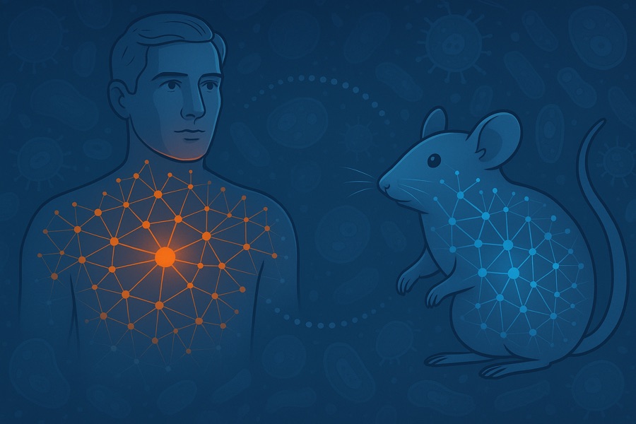
Turning human data into breakthrough drugs
Investing in programmable cell-interaction communities over genes, cells, and “virtual cells.”
The next wave of bispecific antibodies & antibody-mediated vaccines
Built an AI-enabled spatial biology platform identifying immune cell organizational patterns in tumors; led a 5-person cross-functional team; designed a bispecific antibody program targeting a dendritic cell–T cell resistance axis; built clinical-center collaborations across the US, including a paid engagement for the platform's AI-generated insight itself; selected for the Merck Digital Sciences Studio.
Designs and deploys production agentic AI systems inside biotech operations, at Voyant Bio and for other founders and operators, spanning business operations and intelligence, target discovery, therapeutic design, and data analysis. Writes about agentic AI for the operators putting these systems to work.
Writes a biotechnology Substack with 4,300+ subscribers, publishing investment-oriented perspectives on drug development strategy, clinical differentiation, and therapeutic modalities across oncology and immunology.
Conducted scientific diligence on early-stage AI and oncology platforms; built investment theses linking scientific mechanism, platform differentiation, and commercial opportunity; developed scenario-based risk/reward assessments.
Directed translational immunology programs across a 50+ scientist team as the primary bridge between academic research and Regeneron's industrial drug development. Led the program published in Nature Medicine, showing that intratumoral dendritic cell–CD4⁺ T helper cell niches drive the CD8⁺ T cell differentiation behind response to PD-1 blockade, a generalized mechanism across cancer types where these three cell types organize into spatial "triads" that separate responders from non-responders. Built analysis pipelines integrating spatial and single-cell genomics across 10M+ clinical-trial cells.
Pioneered single-cell genomics approaches defining mechanisms of immunotherapy response and resistance; developed ML-based computational frameworks mapping gene interaction networks. Ph.D. 2016–2019 (GPA 4.9/5); recipient of the Research Excellence Award.
Investment-oriented perspectives on drug development strategy, clinical differentiation, and therapeutic modalities, written from having built, invested in, and evaluated these companies directly.
Read Bio Signal